Arnaud G. L'Huillier, Switzerland
Geneva University Hospitals
Epstein - Barr virus driving post-transplant lymphoproliferative disorders: experience of the Swiss Pediatric Liver Center
Nathalie M. Rock1, Antoine Bouroumeau2, Danai Papangelopoulou3,4, Ismini Mainta5, Eirini Katirtzidou6, Barbara E. Wildhaber 1, Arnaud G. L’Huillier 1, Marc Ansari 3,4, Valérie A. McLin 1, Anne-Laure Rougemont 1,2, Frederic Baleydier3,4.
1Swiss Pediatric Liver Center, Department of Pediatrics, Gynecology, and Obstetrics, University Hospitals Geneva and University of Geneva, , Geneva, Switzerland; 2Division of clinical pathology, University Hospitals Geneva and University of Geneva, , Geneva, Switzerland; 3Division of Pediatric Oncology and Hematology, Department of Pediatrics, Gynecology, and Obstetrics, University Hospitals Geneva and University of Geneva, Geneva, Switzerland; 4CANSEARCH research laboratory, Medical Faculty, Geneva University, Geneva, Switzerland; 5Division of Nuclear Medicine, University Hospitals Geneva, Geneva, Switzerland; 6Unit of Pediatric Radiology, Division of Radiology, University Hospitals Geneva, Geneva, Switzerland
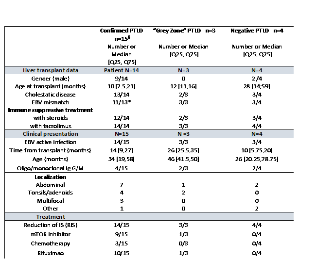
Introduction: Post-Transplant Lymphoproliferative Disorders (PTLD) are a well-known complication of pediatric solid organ transplantation (SOT) which may significantly impair outcomes. EBV is often responsible for triggering PTLD, highlighting the decisive role of the immune system in the development of these disorders. The aim of this study was to describe the diagnostic approach and treatment in a single pediatric liver transplant (pLT) cohort.
Methods: All patients followed in our pLT center who presented with 2 persistently positive EBV DNA in plasma despite reduction of immunosuppression (RIS), were systematically included and evaluated for PTLD according to our local protocol. The following variables were recorded: Symptoms, laboratory parameters, diagnostic approach, choice of treatment, outcome (rejection, infection, loss of graft and death). All cases were retrospectively reviewed by two pathologists following the diagnostic criteria published in the revised 4th edition of the World Health Organization (WHO) Classification in 2017. Three groups were subsequently defined: (1) histologically confirmed PTLD (2) “grey zone” PTLD if all histological criteria were not formally met and (3) no PTLD.
Results: From 2009 to 2021, 21 patients met the inclusion criteria were explored among the 112 patients that had undergone pLT. Patient characteristics, results and treatment approaches are summarized in the Table below. The most common clinical manifestations were general malaise, impaired weight gain, and diarrhea. Seventeen (17/21) patients were evaluated with a total body position emission tomography computed tomography (PET CT) at diagnosis. All patient had a biopsy driven by imaging. Fifteen PTLD in 14 patients were included in group 1. The incidence of PTLD in the overall cohort was 12.5% (14/112). Three further patients were classified in group 2“. Four (4) patients were finally negative for PTLD and included in group 3. The most common subtype of PTLD was non-destructive PTLD (8/15), followed by monomorphic (4/15) and polymorphic PTLD (3/15). In the monomorphic group, 3 patients (3/4) presented with different patterns of PTLD depending on the site of the biopsy or within the same histological specimen.
In 5 patients, including the 3 treated by chemotherapy, liver rejection was observed within 12 months of PTLD diagnosis. Only one PTLD relapse was observed. All patients are alive with their first graft.
Conclusion: A systematic approach for diagnosis and treatment of PTLD including careful histological analysis according to the most recent WHO criteria, results in favorable outcomes. Histological analysis may be challenging in the diagnostic process with very heterogeneous patterns, but remains the cornerstone of the management. Cases remaining in a histological “grey zone” may need treatment decision relying on the patient’s overall condition.
Lectures by Arnaud G. L'Huillier
| When | Session | Talk Title | Room |
|---|---|---|---|
|
Mon-27 08:00 - 09:30 |
COVID in pediatric transplantation | COVID vaccination in pediatric patients | Zilker 3-4 |
|
Tue-28 08:00 - 09:00 |
Infectious Diseases | Infectious Diseases in the First Year After Solid Organ Transplantation in children within the Swiss Transplant Cohort Study | Hill Country AB |
|
Tue-28 09:10 - 10:10 |
Combined Topics | Epstein - Barr virus driving post-transplant lymphoproliferative disorders: experience of the Swiss Pediatric Liver Center | Hill Country CD |